| THE GRAND SUBVERSION, AUTOPSY REPORT RESULTS JFK KENNEDY JOHN F ASSASSINATION : FILE, ( Documents and Reports. ) |
|
[
HOME ]
|
AUTOPSY REPORT RESULTS JFK ASSASSINATION :
Description of the Brain.
( Warren Commission )
General Description of the Body
The body is that of a muscular, well-developed and well nourished adult
Caucasian male measuring 72 1/2 inches and weighing approximately 170 pounds.
There is beginning rigor mortis, minimal dependent livor mortis of the dorsum,
and early algor mortis. The hair is reddish brown and abundant, the eyes are
blue, the right pupil measuring 8 mm. in diameter, the left 4 mm. There is edema
and ecchymosis of the inner canthus region of the left eyelid measuring
approximately 1. 5 cm. in greatest diameter. There is edema and ecchymosis
diffusely over the right supra-orbital ridge with abnormal mobility of the
underlying bone. (The remainder of the scalp will be described with the skull.)
There is clotted blood on the external ears but otherwise the ears, nares, and
mouth are essentially unremarkable. The teeth are in excellent repair and there
is some pallor of the oral mucous membrane.
Situated on the upper right posterior thorax just above the upper border of the
scapula there is a 7 x 4 millimeter oval wound. This wound is measured to be 14
cm. from the tip of the right acromion process and 14 cm. below the tip of the
right mastoid process.
Situated in the low anterior neck at approximately the level of the third and
fourth tracheal rings is a 6. 5 cm. long transverse wound with widely gaping
irregular edges. (The depth and character of these wounds will be further
described below.)
Situated on the anterior chest wall in the nipple line are bilateral 2 cm. long
recent transverse surgical incisions into the subcutaneous tissue. The one on
the left is situated 11 cm. cephalad to the nipple and the one on the right 8
cm. cephalad to the nipple. There is no hemorrhage or ecchymosis associated with
these wounds. A similar clean wound measuring 2 cm. in length is situated on the
antero-lateral aspect of the left mid arm. Situated on the antero-lateral aspect
of each ankle is a recent 2 cm. transverse incision into the subcutnaeous
tissue.
There is an old well healed 8 cm. McBurney abdominal incision. Over the lumbar
spine in the midline is an old, well healed 15 cm. scar. Situated on the upper
antero-lateral aspect of the right thigh is an old, well healed 8 cm. scar.
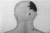
Missile Wounds
There is a large irregular defect of the scalp and skull on the right involving
chiefly the parietal bone but extending somewhat into the temporal and occipital
regions. In this region there is an actual absence of scalp and bone producing a
defect which measures approximately 13 cm. in greatest diameter.
From the irregular margins of the above scalp defect tears extend in stellate
fashion into the more or less intact scalp as follows:
From the right inferior temporo-parietal margin anterior to the right ear to a
point slightly above the tragus.
From the anterior parietal margin anteriorly on the forehead to approximately 4
cm. above the right orbital ridge.
From the left margin of the main defect across the midline antero-laterally for
a distance of approximately 8 cm.
From the same starting point as c. 10 cm. postero-laterally.
Situated in the posterior scalp approximately 2. 5 cm. laterally to the right
and slightly above the external occipital protuberance is a lacerated wound
measuring 15 x 6 mm. In the underlying bone is a corresponding wound through the
skull which exhibits beveling of the margins of the bone when viewed from the
inner aspect of the skull.
Clearly visible in the above described large skull defect and exuding from it is
lacerated brain tissue which on close inspection proves to represent the major
portion of the right cerebral hemisphere. At this point it is noted that the
falx cerebri is extensively lacerated with disruption of the superior saggital
sinus.
Upon reflecting the scalp multiple complete fracture lines are seen to radiate
from both the large defect at the vertex and the smaller wound at the occiput.
These vary greatly in length and direction, the longest measuring approximately
19 cm. These result in the production of numerous fragments which vary in size
from a few millimeters to 10 cm. in greatest diameter.
The complexity of these fractures and the fragments thus produced tax
satisfactory verbal description and are better appreciated in photographs and
roentgenograms which are prepared.
The brain is removed and preserved for further study following formalin
fixation.
Received as separate specimens from Dallas, Texas are three fragments of skull
bone which in aggregate roughly approximate the dimensions of the large defect
described above. At one angle of the largest of these fragments is a portion of
the perimeter of a roughly circular wound presumably of exit which exhibits
beveling of the outer aspect of the bone and is estimated to measure
approximately 2. 5 to 3. 0 cm. in diameter. Roentgenograms of this fragment
reveal minute particles of metal in the bone at this margin. Roentgenograms of
the skull reveal multiple minute metallic fragments along a line corresponding
with a line joining the above described small occipital wound and the right
supra-orbital ridge. From the surface of the disrupted right cerebral cortex two
small irregularly shaped fragments of metal are recovered. These measure 7 x 2
mm. and 3 x 1 mm. These are placed in the custody of Agents Francis X. O'Neill,
Jr. and James W. Sibert, of the Federal Bureau of Investigation, who executed a
receipt therefor (attached).
The second wound presumably of entry is that described above in the upper right
posterior thorax. Beneath the skin there is ecchymosis of subcutaneous tissue
and musculature. The missile path through the fascia and musculature cannot be
easily proved. The wound presumably of exit was that described by Dr. Malcolm
Perry of Dallas in the low anterior cervical region. When observed by Dr. Perry
the wound measured "a few millimeters in diameter", however it was extended as a
tracheostomy incision and thus its character is distorted at the time of
autopsy. However there is considerable eccymosis of the strap muscles of the
right side of the neck and of the fascia about the trachea adjacent to the line
of the tracheostomy wound. The third point of reference in connecting these two
wounds is in the apex (supra-clavicular portion) of the right pleural cavity. In
this region there is contusion of the parietal pleura and of the extreme apical
portion of the right upper lobe of the lung. In both instances the diameter of
contusion and ecchymosis at the point of maximal involvement measures 5 cm. Both
the visceral and parietal pleura are intact overlying these areas of trauma.
Incisions
The scalp wounds are extended in the coronal plane to examine the cranial
content and the customary (Y) shaped incision is used to examine the body
cavities.
Thoracic Cavity
The bony cage is unremarkable. The thoracic organs are in their normal positions
are relationships and there is no increase in free pleural fluid. The above
described area of contusion in the apical portion of the right pleural cavity is
noted.
Lungs
The lungs are of essentially similar appearance the right weighing 320 Gm., the
left 290 Gm. The lungs are well aerated with smooth glistening pleural surfaces
and gray-pink color. A 5 cm. diameter area of purplish red discoloration and
increased firmness to palpation is situated in the apical portion of the right
upper lobe. This corresponds to the similar area described in the overlying
parietal pleura. Incision in this region reveals recent hemorrhage into
pulmonary parenchyma.
Heart
The pericardial cavity is smooth walled and contains approximately 10 cc. of
straw-colored fluid. The heart is of essentially normal external contour and
weighs 350 Gm. The pulmonary artery is opened in situ and no abnormalities are
noted. The cardiac chambers contain moderate amounts of postmortem clotted
blood. There are no gross abnormalities of the leaflets of any of the cardiac
valves. The following are the circumferences of the cardiac valves: aortic 7. 5
cm., pulmonic 7 cm., tricuspid 12 cm., mitral 11 cm. The myocardium is firm and
reddish brown. The left ventricular myocardium averages 1. 2 cm. in thickness,
the right ventricular myocardium 0. 4 cm. The coronary arteries are dissected
and are of normal distribution and smooth walled and elastic throughout.
Abdominal Cavity
The abdominal organs are in their normal positions and relationships and there
is no increase in free peritoneal fluid. The vermiform appendix is surgically
absent and there are a few adhesions joining the region of the cecum to the
ventral abdominal wall at the above described old abdominal incisional scar.
Skeletal System
Aside from the above described skull wounds there are no significant gross
skeletal abnormalities.
Photography
Black and white and color photographs depicting significant findings are exposed
but not developed. These photographs were placed in the custody of Agent Roy E.
Kellerman of the U. S. Secret Service, who executed a receipt therefore
(attached).
Roentgenograms
Roentgenograms are made of the entire body and of the separately submitted three
fragments of skull bone. These are developed are were placed in the custody of
Agent Roy H. Kellerman of the U. S. Secret Service, who executed a receipt
therefor (attached).
Summary
Based on the above observations it is our opinion that the deceased died as a
result of two perforating gunshot wounds inflicted by high velocity projectiles
fired by a person or persons unknown. The projectiles were fired from a point
behind and somewhat above the level of the deceased. The observations and
available information do not permit a satisfactory estimate as to the sequence
of the two wounds.
The fatal missile entered the skull above and to the right of the external
occipital protuberance. A portion of the projectile traversed the cranial cavity
in a posterior-anterior direction (see lateral skull roentgenograms) depositing
minute particles along its path. A portion of the projectile made its exit
through the parietal bone on the right carrying with it portions of cerebrum,
skull and scalp. The two wounds of the skull combined with the force of the
missile produced extensive fragmentation of the skull, laceration of the
superior saggital sinus, and of the right cerebral hemisphere.
The other missile entered the right superior posterior thorax above the scapula
and traversed the soft tissues of the supra-scapular and the supra-clavicular
portions of the base of the right side of the neck. This missile produced
contusions of the right apical parietal pleura and of the apical portion of the
right upper lobe of the lung. The missile contused the strap muscles of the
right side of the neck, damaged the trachea and made its exit through the
anterior surface of the neck. As far as can be ascertained this missile struck
no bony structures in its path through the body.
In addition, it is our opinion that the wound of the skull produced such
extensive damage to the brain as to preclude the possibility of the deceased
surviving this injury. A supplementary report will be submitted following more
detailed examination of the brain and of microscopic sections. However, it is
not anticipated that these examinations will materially alter the findings.
/s/
J. J. HUMES
CDR, MC, USN (497831)
/s/
"J" THORNTON BOSWELL
CDR, MC, USN (489878)
/s/
PIERRE A. FINCK
LT COL, MC, USA
(04-043-322)
Autopsy Report JFK President John F Kennedy.
( Warren Commission )
Gross Description of the Brain
Following formalin fixation the brain seighs 1500 gms. The right cerebral
hemisphere is found to be markedly disrupted. There is a longitudinal laceration
of the right hemisphere which is para-sagittal in position approximately 2. 5
cm. to the right of the of the midline which extends from the tip of the
occipital lobe posteriorly to the tip of the frontal lobe anteriorly. The base
of the laceration is situated approximately 4. 5 cm. below the vertex in the
white matter. There is considerable loss of cortical substance above the base of
the laceration, particularly in the parietal lobe. The margins of this
laceration are at all points jagged and irregular, with additional lacerations
extending in varying directions and for varying distances from the main
laceration. In addition, there is a laceration of the corpus callosum extending
from the genu to the tail. Exposed in this latter laceration are the interiors
of the right lateral and third ventricles.
When viewed from the vertex the left cerebral hemisphere is intact. There is
marked engorgement of meningeal blood vessels of the left temporal and frontal
regions with considerable associated sub-arachnoid hemorrhage. The gyri and
sulci over the left hemisphere are of essentially normal size and distribution.
Those on the right are too fragmented and distorted for satisfactory
description.
When viewed from the basilar aspect the disruption of the right cortex is again
obvious. There is a longitudinal laceration of the mid-brain through the floor
of the third ventricle just behind the optic chiasm and the mammillary bodies.
This laceration partially communicates with an oblique 1. 5 cm. tear through the
left cerebral peduncle. There are irregular superficial lacerations over the
basilar aspects of the left temporal and frontal lobes.
In the interest of preserving the specimen coronal sections are not made. The
following sections are taken for microscopic examination:
From the margin of the laceration in the right parietal lobe.
From the margin of the laceration in the corpus callosum.
From the anterior portion of the laceration in the right frontal lobe.
From the contused left fronto-parietal cortex.
From the line of transection of the spinal cord.
From the right cerebellar cortex.
From the superficial laceration of the basilar aspect of the left temporal lobe.
JFK ASSASSINATION JOHN F KENNEDY PHOTOS PICTURES
ASSASSIN WHO KILLED AUTOPSY REPORT RESULTS.
JFK AUTOPSY KENNEDY REPORT RESULTS JOHN F ASSASSINATION
CONSPIRACY PHOTOS PICTURES.
JFK John F Kennedy Assassination:
[
HOME ]
![]()
JFK AUTOPSY PAGE (5) [
BACK
]
![]()
SECTIONS
[Parkland Hospital]--[Bethesda
Autopsy]--[Dallas I]--[Dallas
II]--[Shots]--[Magic
Single Bullet Theory]--[Oswald
I]--[Oswald II]--[Oswald
III]--[Oswald
IV]--[Three
Tramps]--[Dallas
Police]
|
Page Subject : List of Internal Links. |
||
|
Grand Subversions Links |
MEDICAL - HEALTH LINKS. | |
|
Grand Subversions JFK Assassination VIEW ALL SECTIONS -[ PHOTO LINK INDEX ] |
|
Parkland Hospital PAGES 1 - 4 |
Bethesda Autopsy PAGES 1 - 5 | ||||
 |
 |
 |
 |
 |
 |
|
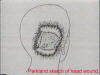
1
Arrival and treatment at Parkland Hospital. 2 Parkland description of head wound vs Autopsy 3 THROAT WOUND 4 THE BODY SNATCHERS MORE IN AUTOPSY SECTION. |
1
AUTOPSY PHOTOS 2 THE SERIOUS PROBLEMS 3 Dallas Doctors Testimony, HEAD and THROAT WOUND |
4
The Back Wound and the finding of the
Magic Bullet 5 Presidents clothing, x-rays, Dallas Doctors view Autopsy Photos AFTER 25 YEARS |
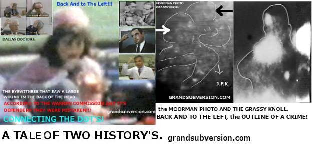
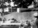
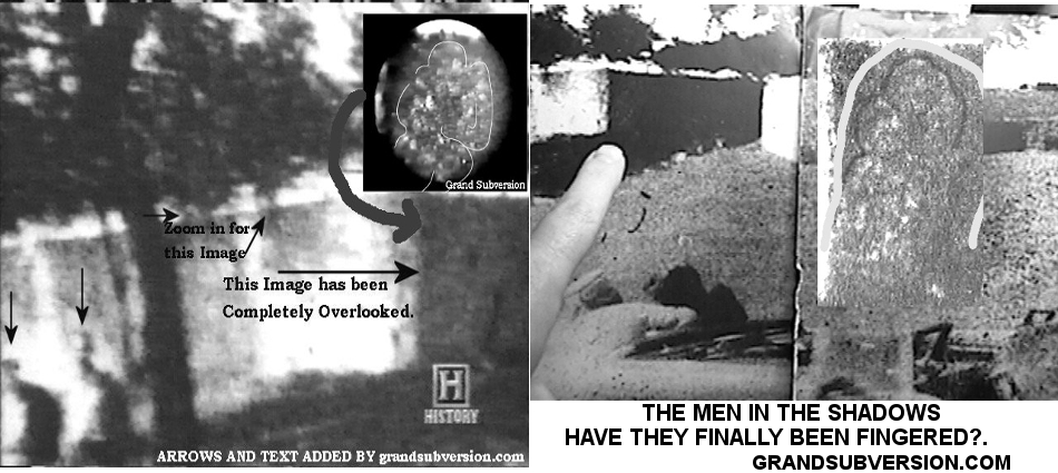
| grandsubversions.com NEW Grassy Knoll ENHANCEMENTS. | |||
| Moorman photo. | Black Dog Image. | Knoll Overview. | Gordon Arnold Factor. |
 |
 |
 |
 |
| CLICK IMAGE FOR ENHANCEMENT PAGE. |
JFK John F Kennedy Assassination:
[
HOME ]
![]() JFK Autopsy PAGE (5)
[
BACK
]
JFK Autopsy PAGE (5)
[
BACK
]
![]()

JFK AUTOPSY REPORTS RESULTS DOCTORS FILM VIDEO HEAD WOUND KENNEDY ASSASSINATION
PHOTOS.
AUTOPSY REPORT RESULTS JFK JOHN F KENNEDY ASSASSINATION PHOTOS PICTURES INJURY
WOUND CONSPIRACY
CRIME SCENE PHOTOS COLD CASE JFK FILES CSI DALLAS POLICE WHO KILLED KENNEDY RFK
MLK JR.ASSASSINATION
.
JFK AUTOPSY
SECTION. PAGE (1)
JFK AUTOPSY REPORT RESULTS KENNEDY ASSASSINATION COVER UP CONSPIRACY PHOTO
PICTURES THEORIES.
autopsy photos report pictures jfk kennedy jr robert wounds headshot grassy
knoll doctors assassination dallas police shooting
JFK John F Kennedy Assassination
CONSPIRACY:
[
HOME ]
![]()